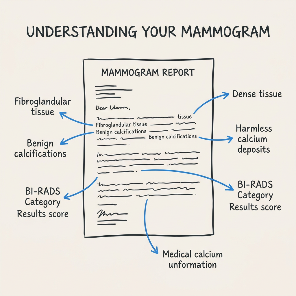
You opened the letter from your mammogram center, and somewhere between the “normal results” line and the bottom of the page, there it was: “You have dense breast tissue.”
So you did what most of us do. You googled it. And now you’re reading about cancer risk and BI-RADS categories and supplemental screening, and it all feels like a lot more than you expected from a routine mammogram.
Take a breath. About half of all women who get mammograms receive this same letter. Dense breast tissue is common, it is not a diagnosis, and there are straightforward things you can do with this information. Let’s walk through it.
What Dense Breast Tissue Actually Is
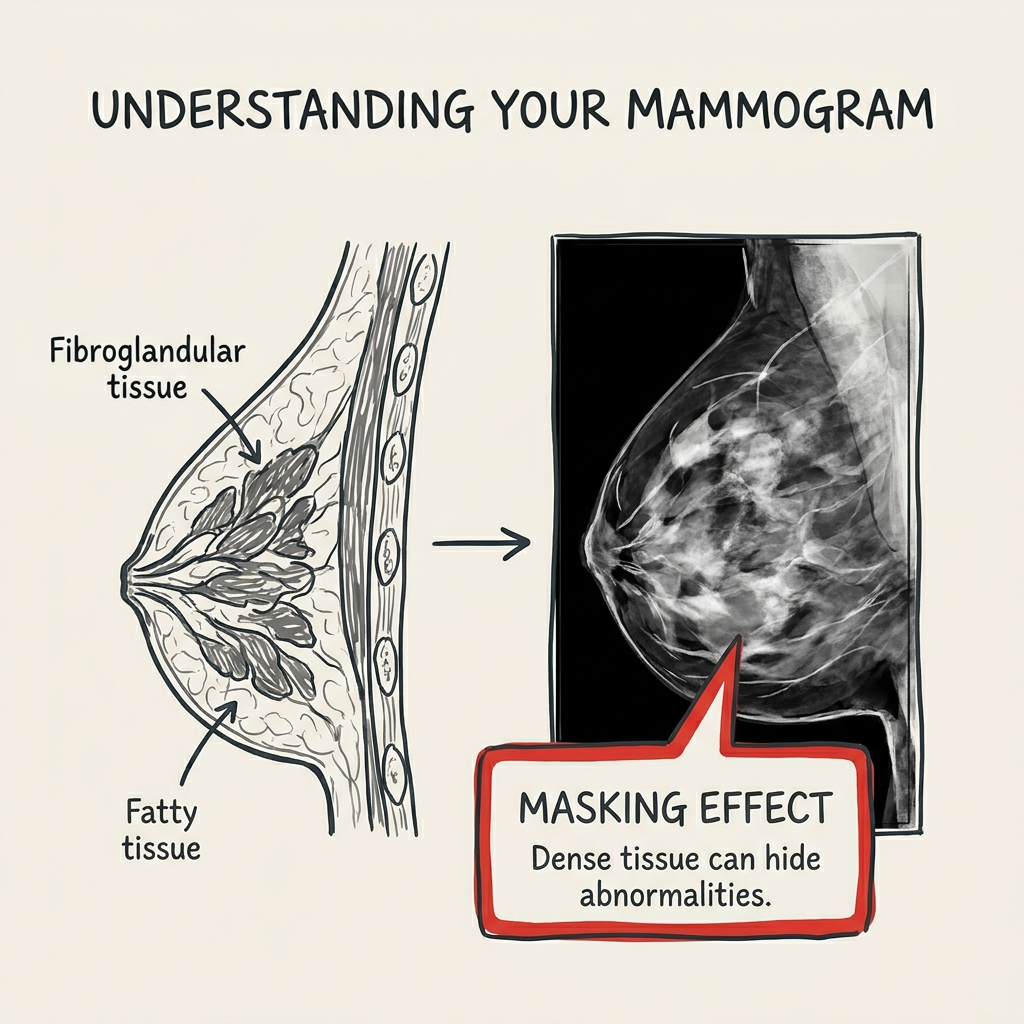
Your breasts are made of three types of tissue: fatty tissue, fibrous connective tissue, and glandular tissue (the parts that produce and carry milk). Dense breast tissue means you have more fibrous and glandular tissue than fat.
On a mammogram, dense tissue and cancers both show up white. That is the core concern: dense tissue can make small tumors harder to spot. Not because density is dangerous on its own, but because it can hide things.
Key takeaway: Dense breast tissue means more fibrous and glandular tissue than fat. It is common, not a diagnosis, and the main concern is that it can make mammograms harder to read.
The Four Density Categories
Radiologists classify breast density into four levels, using a system called BI-RADS (Breast Imaging Reporting and Data System):
A. Almost entirely fatty. Mammograms are very easy to read. About 10% of women fall here.
B. Scattered fibroglandular density. Some dense tissue, but mostly fatty. Mammograms are still fairly clear. About 40% of women.
C. Heterogeneously dense. More dense tissue than fatty. This is the category that catches many women off guard because it is the most common “dense” classification. About 40% of women.
D. Extremely dense. Almost entirely dense tissue. Mammograms are the most difficult to read. About 10% of women.
If your letter said “heterogeneously dense,” you are in the largest group. Categories C and D are both considered “dense,” and together they account for roughly half of all women screened.
Your density is not something you caused or can change through diet or exercise. It is influenced by genetics, age, hormone levels, and body composition. Density often decreases after menopause, but not always.
Key takeaway: Categories C and D are both “dense.” About half of all women fall into one of these two groups.
Does Dense Breast Tissue Increase Cancer Risk?
This is the question that sends most women down a rabbit hole after reading the letter. The honest answer: slightly, and with important context.
Women with dense breast tissue (categories C and D) have a modestly higher risk of developing breast cancer compared to women with fatty breasts. But “higher risk” does not mean “high risk.” Density is one factor among many, alongside family history, age, genetic markers, and lifestyle. Most women with dense breasts will never develop breast cancer.
The bigger practical concern is the masking effect. Dense tissue makes small cancers harder to see on a standard mammogram. Your doctor may recommend additional screening not because you are in immediate danger, but because getting a clearer picture is a reasonable step.
If you are curious about how genetic factors play into breast cancer risk beyond density, understanding markers like BRCA can help put the pieces together.
Key takeaway: Dense breast tissue slightly increases cancer risk, but the bigger issue is that it can hide cancers on a standard mammogram. Additional screening helps close that gap.
What Your Doctor Might Recommend Next
If your mammogram shows dense tissue, your doctor has a few tools to get a clearer view:
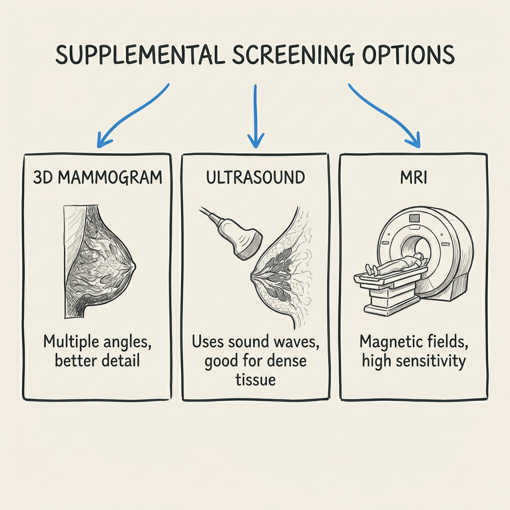
3D mammogram (tomosynthesis). Takes multiple X-ray images from different angles to create a layered picture. Better at finding cancers in dense tissue than a standard 2D mammogram. Many screening centers already use it as the default.
Breast ultrasound. Can find cancers that mammograms miss in dense tissue. Painless, no radiation, and often recommended for women in categories C and D.
Breast MRI. The most sensitive screening tool available, but typically reserved for women with additional risk factors beyond density alone, such as a strong family history or a known genetic mutation.
Bring the letter to your next appointment and ask your doctor which option, if any, fits your situation. The conversation takes five minutes and gives you a clear next step.
Breast self-examination is another way to stay familiar with how your breasts normally feel, so you notice changes between mammograms.
Key takeaway: 3D mammography and breast ultrasound are the most common supplemental screening options for dense breast tissue. Ask your doctor which one fits your risk profile.
Why You Got This Letter Now
If you have been getting mammograms for years and never received a density notification before, it is not because your breasts suddenly changed. In 2024, the FDA finalized a rule requiring all mammography facilities in the United States to notify patients about their breast density. Before that, notification laws varied by state, and many women were never told.
The letter is not a warning. It is information you should have been receiving all along.
Breast density can change over time. Hormonal shifts during menopause, pregnancy, or hormone replacement therapy can all affect density. Your classification at 45 may be different from your classification at 60. That is normal.
Key takeaway: The 2024 FDA rule means mammography centers must now tell you about breast density. This letter is not new information about your body; it is information that is finally being shared.
What to Do with This Information
Dense breast tissue does not require treatment. What it gives you is a piece of the picture that helps you and your doctor make informed decisions about screening.
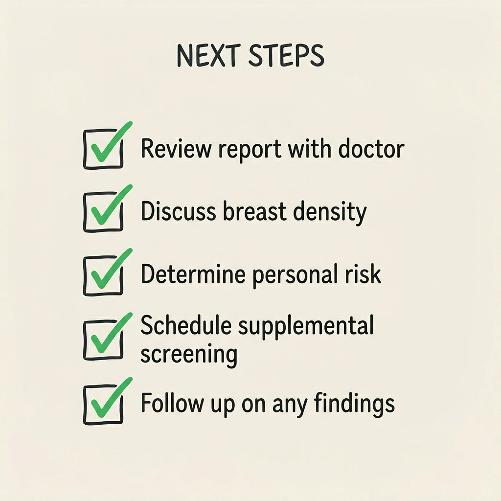
A practical checklist:
- Keep the letter. Bring it to your next doctor’s appointment.
- Ask about supplemental screening. Specifically, ask whether a 3D mammogram or breast ultrasound is appropriate for you.
- Know your other risk factors. Family history, age, genetic mutations, and previous biopsies all factor into your overall risk profile. Your doctor can put density in context with everything else.
- Stay consistent with mammograms. Dense tissue makes screening more important, not less. Do not skip appointments because the results feel confusing.
- Practice self-examination. Knowing your own baseline makes it easier to notice changes.
If additional screening ever finds something that needs a closer look, knowing what to expect from a mastectomy recovery and understanding your surgical options can help you feel less alone in the process. But for most women, the next step is simply a conversation with their doctor.
You Have Someone to Talk To
We know this is not the kind of thing you forget about after closing a browser tab. Even when the medical facts say “this is common” and “this is manageable,” it can still feel heavy.
Front Room Underfashions has been sitting across from women in moments exactly like this one for over 40 years. Lenore Shebuski founded the shop in 1980 after her own breast cancer diagnosis because she believed women deserved better than buying a prosthesis at a medical supply store. Crystal Bliese and her daughter Heather Tynes carry that same belief today at three Lansing-area locations, including two inside cancer centers.
We are not your doctor. But we are the place where women come to ask questions, get a comfortable, certified fitting, and talk to someone who has heard it all and still has time for you.
If you want to come in, come in. If you just want to call and ask a question, do that. We are here for every chapter.
Front Room Underfashions is a certified mastectomy fitting boutique serving Lansing-area women since 1980. With ABC-accredited fitters and three locations, including the Herbert Herman Cancer Center and Karmanos/McLaren Greater Lansing, Front Room helps every woman find the right fit at every stage of life. Schedule a free fitting or call to learn more.